Choose the Appropriate Treatment for Your Dental Needs
Oral surgery is not one procedure. The recommended treatment depends on the condition of the tooth, surrounding bone and gums, nearby anatomy, infection, future restorative needs and your general health. Your dentist will explain whether treatment should involve removal, monitoring, grafting or another approach.
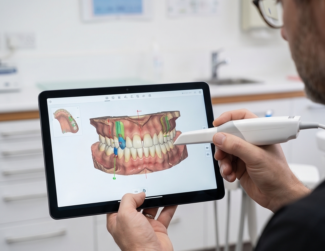
Remove a Tooth Only When Saving It Is No Longer Predictable
Extraction may be recommended when a tooth is severely damaged, fractured, infected or has lost too much support to be restored predictably. Before removing a tooth, we assess whether root canal treatment, restorative treatment or periodontal care could reasonably preserve it. If extraction is required, the procedure is planned with the surrounding gum and bone and any future tooth replacement in mind.
- Removes teeth that cannot be restored predictably
- Treats a source of persistent infection or pain when preservation is no longer appropriate
- Allows the extraction to be planned around surrounding bone and soft tissue
- Creates an opportunity to discuss implant, bridge or denture replacement before treatment
- Socket preservation can be considered when future implant treatment is planned
- Includes personalised recovery and follow-up instructions
Before & After
Real results from patients who underwent Tooth Extraction at our clinic.
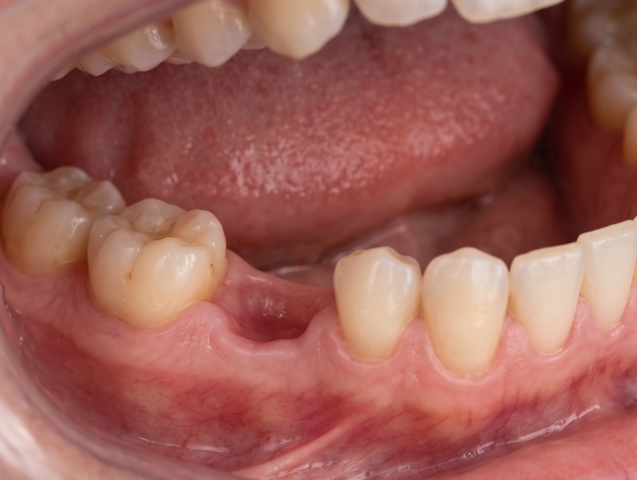
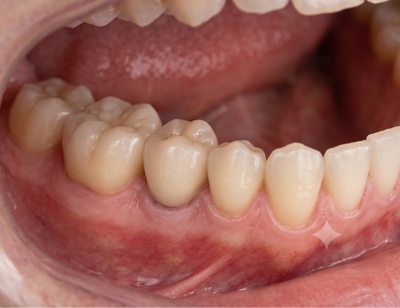
We assess decay, cracks, infection, periodontal support, remaining tooth structure and realistic restorative alternatives.
Appropriate dental imaging helps assess root shape, bone and nearby structures. Future replacement or graft requirements can be discussed before treatment begins.
Local anaesthesia is used to provide comfort before the tooth is removed.
The extraction technique is selected according to the tooth, roots and surrounding tissues. The socket is cleaned and sutures or graft material are used only when clinically appropriate.
You receive written guidance about clot protection, cleaning, eating, medication and symptoms that should prompt a review.
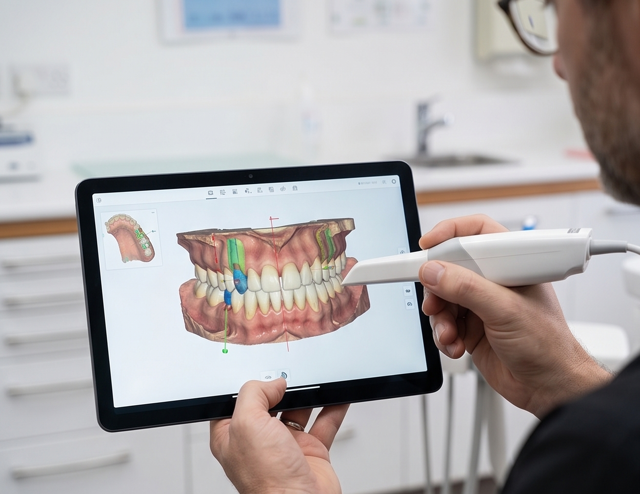
Treat Problematic Wisdom Teeth With Careful Planning
Wisdom teeth do not automatically need to be removed. Extraction may be recommended when a wisdom tooth is causing repeated infection, pain, decay, food trapping, gum problems or damage to the neighbouring tooth. For impacted or partially erupted wisdom teeth, dental imaging helps assess tooth position, root anatomy and nearby structures before treatment.
- Treats repeated inflammation or infection around problematic wisdom teeth
- Reduces food trapping in areas that cannot be cleaned adequately
- Helps protect the neighbouring second molar when it is being affected
- Assesses impacted tooth position before surgery
- Allows nerve proximity and root anatomy to be evaluated where relevant
- Provides practical recovery advice for work, travel and everyday activities
Before & After
Real results from patients who chose Wisdom Teeth Removal at our clinic.
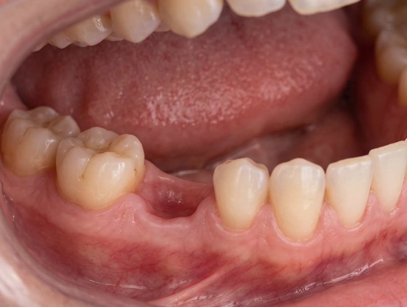
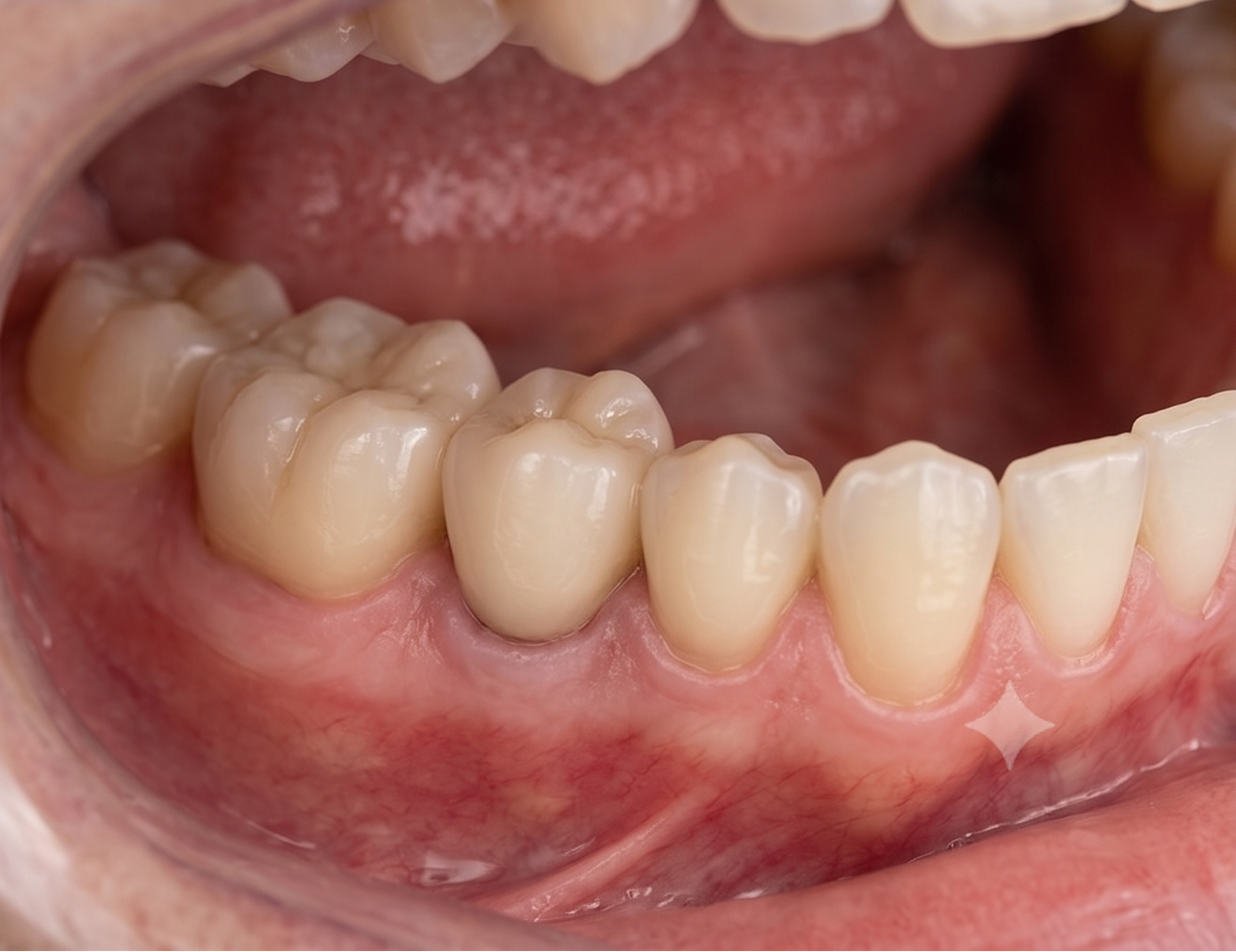
We assess pain, swelling, repeated infections, decay, gum condition and effects on neighbouring teeth.
A panoramic X-ray and CBCT when clinically indicated help evaluate impaction, root shape and nearby anatomy.
Local anaesthesia is planned according to the procedure, and any appropriate additional comfort options are discussed.
When required, a small opening in the gum or surrounding bone allows the tooth to be removed safely. The area is cleaned and sutured when appropriate.
You receive clear instructions about swelling, oral hygiene, diet, clot protection and symptoms that require review.

Rebuild Bone for Future Tooth Replacement
Jawbone naturally changes after tooth loss, infection or trauma. In some areas, there may not be enough bone volume to support an implant in an appropriate position. Dental bone grafting is used to preserve or rebuild selected areas of the jaw when additional bone support is required for future treatment. The type and timing of grafting depend on the size of the defect, soft tissues, implant plan and individual healing factors.
- Preserves selected extraction sites when future implant treatment is planned
- Rebuilds local bone deficiencies
- Helps create adequate support for implant positioning
- Can improve the shape of the ridge where bone volume has reduced
- Uses graft type and technique according to the individual defect
- Allows implant placement to be staged until healing is sufficient
Before & After
Real results from patients who chose Bone Grafting at our clinic.
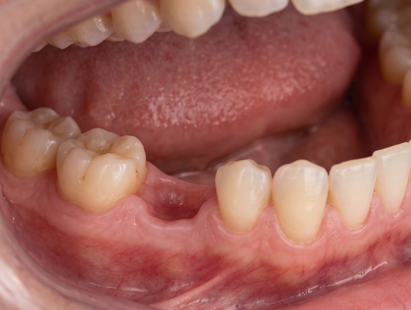
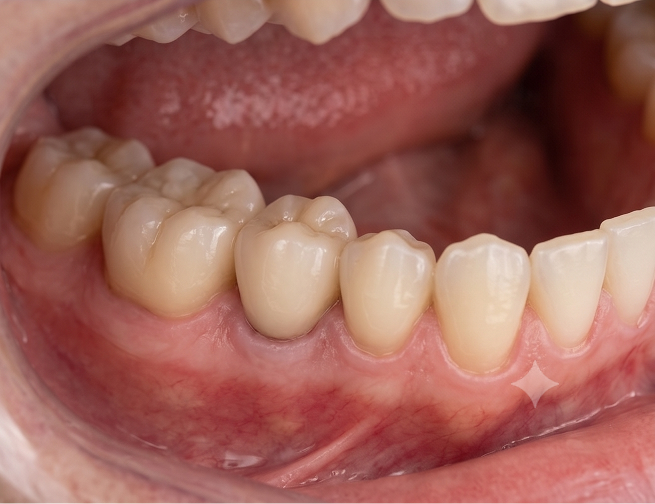
Clinical examination and appropriate 3D imaging evaluate bone width, height, soft tissues and nearby anatomy.
The graft is designed according to the intended implant and final restoration rather than simply adding bone without a restorative plan.
The surgical area is prepared and graft material is placed and stabilised. A membrane or fixation may be used when required.
Sutures, cleaning instructions and careful temporary-restoration design help minimise unnecessary pressure on the graft.
Healing is evaluated clinically and with additional imaging when appropriate before the next treatment stage is started.
Create Bone Support for Implants in the Upper Back Jaw
After upper back teeth are lost, the remaining bone beneath the maxillary sinus may be insufficient for implant placement. A sinus lift carefully elevates the sinus membrane and places graft material beneath it to increase available bone height for future implant support. The amount of existing bone and the planned implant position help determine whether a smaller internal approach or a lateral sinus-lift technique is more appropriate.
- Creates additional bone height in selected upper-molar implant sites
- Allows treatment planning around the anatomy of the maxillary sinus
- Uses CBCT imaging to assess existing bone and sinus structures
- Can be performed using different surgical approaches depending on the deficiency
- Implant placement may sometimes be combined with the graft when adequate stability is achievable
- Provides a staged option when implant placement should wait for graft healing
Before & After
Real results from patients who chose Sinus Lift Surgery at our clinic.
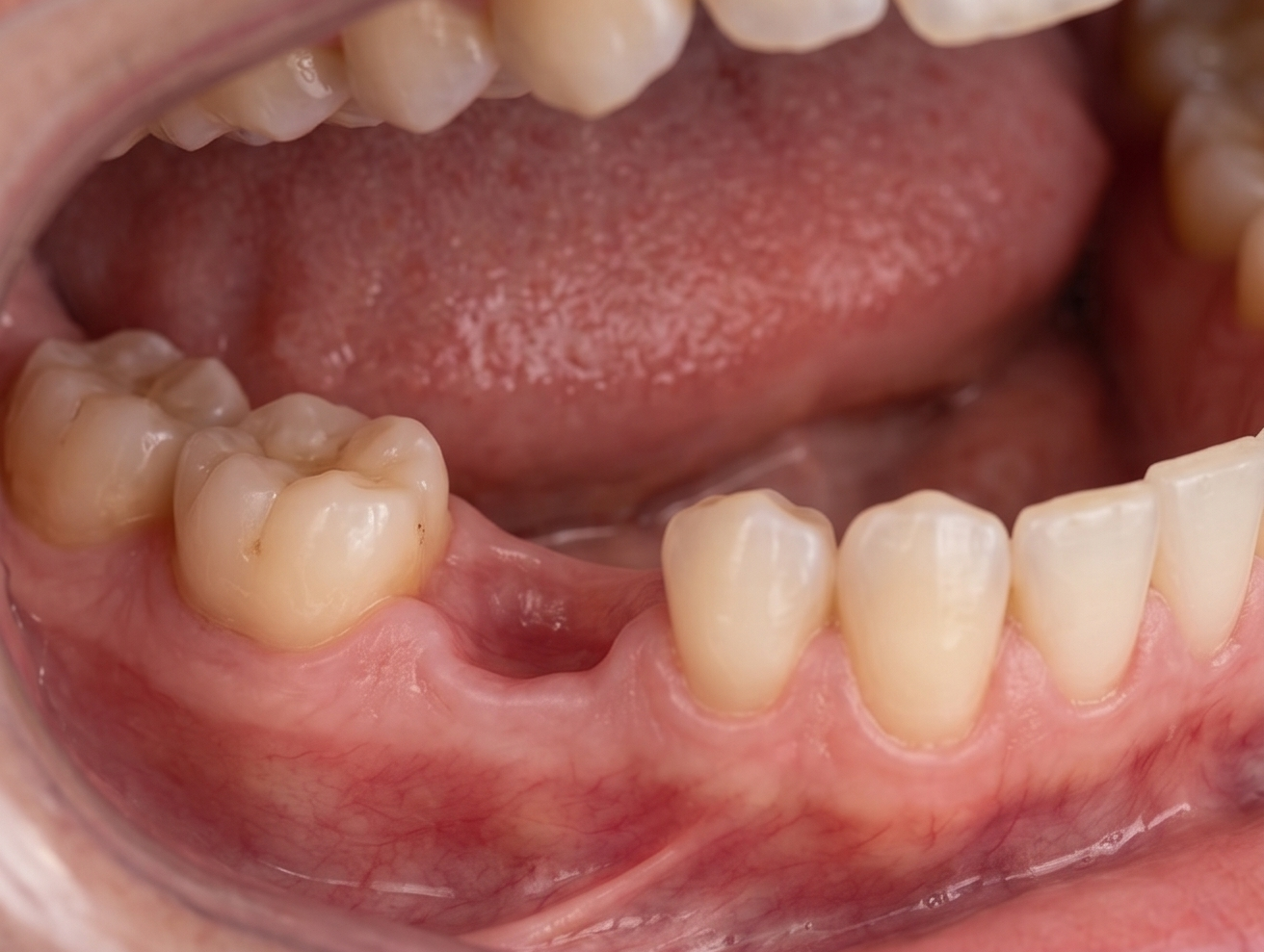
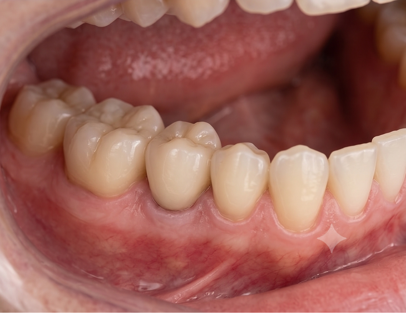
CBCT imaging helps evaluate remaining bone height, sinus anatomy, nearby teeth and the planned implant site.
The amount of bone present and the implant plan determine whether an internal or lateral sinus-lift approach should be considered.
The membrane is carefully raised to create space for graft material.
Graft material is placed beneath the elevated membrane. Implant placement may occur during the same procedure only when the available bone provides sufficient initial stability.
You receive sinus-specific recovery instructions and follow-up before the implant is restored or the next treatment stage begins.